That sudden, throbbing pain in your joint red, hot, and so tender a bedsheet feels like agony can be a frightening and confusing experience. Is it a simple injury, a serious infection, or something else entirely For millions, this unmistakable constellation of symptoms points to gout, a form of inflammatory arthritis that demands specific knowledge and action. The challenge lies in accurately identifying it. By the end of this guide, you will be equipped with a clear understanding of gout’s unique signature. You will be able to compare your symptoms against the classic hallmarks of a gout attack and, crucially, know how to distinguish it from other dangerous conditions. Most importantly, you will have the confidence to decide when it’s time to manage your symptoms at home and when that swelling is a red flag requiring urgent medical help.
how to differentiate gout-related swelling from other types of swelling
This requires a clear, practical guide focusing on distinctive features. The previous conversation established context about gout triggers, so I can build on that.
I should structure this to help the user identify key characteristics of gout swelling versus other conditions. The response needs to cover visual cues, pain patterns, timing, and risk factors. A comparison table would be useful for quick reference, but the main focus should be on gout’s unique presentation – like the sudden severe pain in specific joints.
It’s also important to emphasize the need for professional diagnosis since symptoms can overlap with other conditions. The user likely wants actionable knowledge to decide when to seek medical help. I’ll include clear red flags that warrant immediate doctor consultation.
Of course. Knowing if swelling is caused by gout involves looking for a specific set of signs and symptoms that distinguish it from other types of arthritis or inflammation.
What Exactly Is Gout and Why Does It Cause Swelling
Why Gout Attacks Often Start at Night and in the Big Toe
Ever wonder why gout seems to strike while you’re asleep, and usually targets your big toe There’s actually a science behind it. At night, your body temperature drops slightly, and your joints especially those farthest from your heart, like your toes become cooler. Uric acid crystals are more likely to form in cooler temperatures, so the big toe becomes the perfect target. Plus, when you’re lying still, circulation slows down, giving those crystals more time to settle. Combine that with mild dehydration from sleeping for hours without water, and you’ve got the perfect storm for a gout flare-up.
Gout is a common and complex form of inflammatory arthritis characterized by sudden, severe attacks of pain, redness, and tenderness in joints. The root cause is an elevated level of uric acid in the blood, a condition known as hyperuricemia. Uric acid is a normal waste product created when the body breaks down purines, substances found in certain foods and drinks. When uric acid levels become too high, the excess can form microscopic, needle-shaped urate crystals that deposit in a joint or surrounding tissue.
It is the body’s intense inflammatory response to these foreign, sharp crystals that causes the dramatic swelling, heat, and excruciating pain of a gout attack. Essentially, the immune system recognizes the crystals as a threat, launching a full-scale inflammatory assault that results in the classic, debilitating symptoms of gout.
How uric acid crystals trigger inflammation
The process begins when needle-like uric acid crystals deposit in the joint cavity. The immune system doesn’t recognize these sharp crystals as normal substances, but rather as dangerous foreign bodies, much like bacteria. In response, immune cells rush to the site and attempt to engulf the crystals.
This triggers a powerful and complex inflammatory cascade. It activates a protein complex, which in turn signals the release of potent inflammatory chemicals, most of which cause the blood vessels in the area to dilate and leak, resulting in redness and warmth. It also attracts a massive influx of other white blood cells to the joint. This powerful cellular response leads to intense pain, swelling, and tissue inflammation, which characterizes a painful gout attack.
Gout is a common and complex form of inflammatory arthritis characterized by sudden, severe attacks of pain, redness, and tenderness in joints. The root cause is an elevated level of uric acid in the blood, a condition known as hyperuricemia. Uric acid is a normal waste product created when the body breaks down purines, substances found in certain foods and drinks. When uric acid levels become too high, the excess can form microscopic, needle-shaped urate crystals that deposit in a joint or surrounding tissue.
It is the body’s intense inflammatory response to these foreign, sharp crystals that causes the dramatic swelling, heat, and excruciating pain of a gout attack. Essentially, the immune system recognizes the crystals as a threat, launching a full-scale inflammatory assault that results in the classic, debilitating symptoms of gout.
Common joints affected and why the big toe is often first
While gout can strike any joint, it most commonly affects the lower body, particularly the big toe, as well as the ankles, knees, and heels. The reason the base of the big toe is so frequently the first and most classic site of a gout attack is twofold, rooted in basic physics and physiology.
First, as the furthest point from the heart and a relatively exposed area, the big toe is one of the cooler parts of the body. Uric acid is less soluble in cooler temperatures, making it more likely to crystallize out of the blood and into the joint space. Second, the joints in the feet endure significant mechanical stress and minor trauma from walking and bearing our body’s weight. This physical wear and tear is believed to encourage the formation of the sharp, needle-like urate crystals, which then trigger the intense pain, swelling, and inflammation characteristic of a gout flare.
Typical Gout Swelling Symptoms
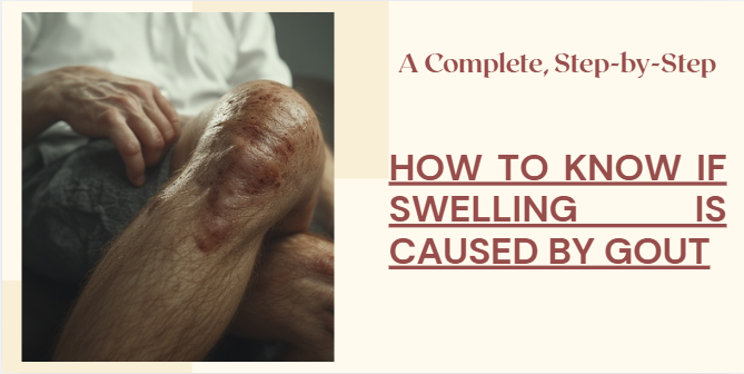
The swelling from a gout attack is characteristically rapid, severe, and unmistakable. Unlike typical joint soreness, it often erupts overnight, transforming a healthy joint into a visibly inflamed and swollen hub of pain within hours. The affected area becomes intensely red, hot, and so tender that even the light weight of a bedsheet can feel unbearable.
The swelling is caused by a fierce inflammatory response as the body tries to attack the urate crystals lodged in the joint, leading to a significant buildup of fluid and white blood cells. This results in a pronounced, often shiny, and distorted appearance to the joint, making movement impossible and marking a classic, debilitating gout flare-up that can last for days or even weeks without treatment.
Timing and onset (why it strikes at night)
Gout’s notoriously sudden and painful onset, often striking in the middle of the night, is not a coincidence but a consequence of several physiological factors converging while you sleep. First, during sleep, your body becomes slightly dehydrated, leading to a higher concentration of uric acid in the bloodstream, which makes crystallization more likely. Second, your core body temperature drops slightly, and this lower temperature, particularly in the extremities like the big toe, further reduces the solubility of uric acid, encouraging the formation of those needle-like crystals.
Finally, while you are at rest, your breathing and circulation slow down, leading to reduced blood flow and a decrease in the anti-inflammatory hormone cortisol. This perfect storm of dehydration, cooler temperatures, and a subdued inflammatory response allows urate crystals to form and trigger a fierce inflammatory reaction by the time you wake.
How the swelling looks and feels
The swelling from a gout attack presents a dramatic and unmistakable picture of intense inflammation. Visually, the affected joint becomes severely swollen and distorted, often losing its natural contour and appearing bulbous. The skin over the joint stretches taut, becoming shiny, and takes on a deep red or purplish hue, similar to a severe infection. To the touch, the area is intensely hot, radiating heat that can often be felt without even making contact.
This heat is a direct result of the inflammatory process. The sensation is one of unbearable, throbbing pain, compounded by a feeling of immense pressure and tightness from within the joint. Even the slightest movement or the gentlest touch, such as from a sock or a bedsheet, can provoke a wave of excruciating pain, making the limb feel utterly useless and exquisitely sensitive.
How long a gout flare usually lasts
The duration of a gout flare can vary, but a typical, untreated attack follows a predictable pattern. An acute gout flare often reaches its peak intensity within 12 to 24 hours of onset. Without any treatment, the severe pain and swelling can persist for anywhere from 5 to 7 days, with milder discomfort and residual inflammation lingering for up to another week or two before fully resolving.
However, with prompt and appropriate anti-inflammatory medication, the duration can be significantly shortened ; a treated flare may be brought under control within a few days. It is crucial to note that while the first flare may subside on its own, subsequent attacks often last longer and affect more joints if the underlying high uric acid levels are not managed, making proper long-term treatment essential.
Gout vs. Other Causes of Swelling
Distinguishing gout from other causes of joint swelling, like osteoarthritis or a bacterial infection, hinges on recognizing its unique onset and symptoms. While an injury causes immediate trauma and osteoarthritis brings a gradual, grinding stiffness, gout strikes with a sudden, explosive intensity, often overnight.
Unlike the diffuse swelling of a sprain, gout typically presents with dramatic, localized signs the skin over the joint becomes intensely red, purplish, and feels noticeably hot to the touch a level of heat less common in routine arthritis. The pain is often described as exquisitely tender, even to the light touch of a bedsheet, which is not typical of standard wear-and-tear arthritis.
While these clues strongly suggest gout, a definitive diagnosis is crucial, as a septic joint (bacterial infection) can mimic some of these signs and requires urgent medical intervention. A doctor can confirm gout by identifying urate crystals in fluid drawn from the swollen joint.
Quick comparison chart (Gout / Pseudogout / Infection / Rheumatoid)
While joint pain and swelling are common to several conditions, key differences in their cause, onset, and pattern help distinguish them. The following comparison outlines the primary characteristics of four common culprits.
- Goutis caused by uric acid crystals (monosodium urate). It typically presents with a sudden, severe attack (often at night), frequently targeting the big toe base. The pain peaks rapidly, and the joint is intensely red, hot, and exquisitely tender. Diagnosis is confirmed by finding needle-shaped, negatively birefringent crystals in joint fluid.
- Pseudogoutis caused by calcium pyrophosphate (CPP) crystals. It mimics gout’s sudden, severe attack but more commonly affects the knee or wrist. The joint is similarly red, hot, and swollen. It is definitively diagnosed by finding rhomboid-shaped, positively birefringent crystals in joint fluid, and X-rays often show cartilage calcification (chondrocalcinosis).
- Septic Arthritis (Infection)is caused by a bacterial infection inside the joint. It causes a sudden, severe hot, swollen, and red joint, often accompanied by fever and chills. It can affect any joint, but commonly the knee. It is a medical emergency diagnosed by finding bacteria and a high white blood cell count in the joint fluid, which requires immediate antibiotics and often surgical drainage.
- Rheumatoid Arthritis (RA)is an autoimmune It presents with a gradual onset of symmetrical stiffness and swelling (e.g., both wrists or both knees). Morning stiffness lasting over 30 minutes is a hallmark. It is not typically associated with the same intense, localized heat as an acute gout attack and is diagnosed through blood tests (e.g., Rheumatoid Factor, anti-CCP) and clinical evaluation.
| Feature | Gout | Pseudogout | Septic Arthritis (Infection) | Rheumatoid Arthritis (RA) |
| Primary Cause | Uric Acid Crystals | Calcium Pyrophosphate (CPP) Crystals | Bacterial Infection | Autoimmune Disorder |
| Typical Onset | Sudden (hours), often at night | Sudden (hours to days) | Sudden (hours) and severe | Gradual (weeks to months) |
| Common Joints | Big toe base, ankle, knee | Knee, wrist, shoulder | Knee, hip, any single joint | Hands, wrists, feet (symmetrical) |
| Key Symptoms | Excruciating pain, intense redness, heat, exquisite tenderness | Severe pain, swelling, redness, heat | Severe pain, swelling, fever, chills | Pain, swelling, prolonged morning stiffness |
| Diagnostic Clue | Needle-shaped crystals in joint fluid | Rhomboid-shaped crystals in joint fluid | Bacteria & high WBC in joint fluid | Positive blood tests (RF, anti-CCP), X-ray changes |
Visual side-by-side image comparison
A visual side-by-side comparison of a gout-affected joint versus a healthy one provides the most immediate and powerful understanding of the condition’s impact. On one side, a healthy joint such as the big toe appears normal in contour and skin color, showing the natural alignment and structure. In stark contrast, the image of an acute gout flare reveals dramatic, localized swelling that distorts the joint’s outline, accompanied by intense redness or purplish discoloration of the skin. The affected area often looks shiny, stretched taut from the inflammation, and visibly warmer in thermal imaging.
This direct visual comparison effectively highlights the sudden, severe, and isolated nature of gout swelling, differentiating it from the more symmetrical swelling of conditions like rheumatoid arthritis or the generalized bruising of an injury. Such imagery is an invaluable tool for both patient education and clinical diagnosis, making the abstract concept of “uric acid crystals” a tangible and recognizable reality.
The Gold Standard: Joint Aspiration and Crystal Analysis
While symptoms can strongly suggest gout, the definitive diagnosis, often called the “gold standard,” is achieved through joint aspiration and crystal analysis. This procedure involves using a needle to draw a small sample of synovial fluid from the inflamed joint. The fluid is then examined under a polarized light microscope.
A positive diagnosis for gout is confirmed by the presence of needle-shaped, negatively birefringent urate crystals within white blood cells. This test is crucial because it not only unequivocally confirms gout but also definitively rules out other dangerous mimics, such as a septic (bacterial) joint infection, or other crystal-induced arthropathies like pseudogout, which requires completely different crystals and treatment. Therefore, joint aspiration provides a level of diagnostic certainty that blood tests and clinical examination alone cannot match.
What happens during the aspiration procedure
A joint aspiration, the procedure used to diagnose gout, is a quick and relatively straightforward in-office test. After cleaning the skin with an antiseptic solution to prevent infection, your doctor will inject a local anesthetic to numb the area, which may cause a brief stinging sensation.
Once the skin is numb, a needle is carefully inserted into the swollen joint to withdraw a sample of the synovial fluid. You may feel a sense of pressure during this part of the procedure. The fluid withdrawal itself typically takes only a minute or two. The extracted fluid, which may appear cloudy or inflamed during a gout attack, is then sent to a lab for immediate analysis under a specialized microscope to identify the presence of urate crystals, providing a definitive diagnosis.
What the lab looks for (crystals under microscope)
In the laboratory, the definitive search for gout hinges on identifying the very culprits that cause the pain urate crystals. The synovial fluid sample is placed on a slide and examined under a polarized light microscope. This specialized microscope uses filters to create a distinctive color pattern, known as birefringence, as light passes through crystals. Technicians are trained to look for specific, tell-tale signs.
the presence of needle-shaped, intracellular crystals inside white blood cells that are strongly negatively birefringent. This means they appear a brilliant yellow when aligned parallel to the microscope’s compensator and blue when perpendicular. Finding these distinctive “needles” inside the cells that have engulfed them is the conclusive evidence of a gout attack, as it directly links the crystals to the inflammatory process. This method also easily distinguishes gout from pseudogout, which features shorter, rhomboid-shaped crystals with weak, positive birefringence.
Why uric acid levels may look normal during a flare
It is a common and often confusing scenario a patient presents with the classic, excruciating symptoms of a gout flare, yet a blood test reveals a normal or only slightly elevated uric acid level. This paradox occurs due to the body’s intense inflammatory response. During an acute attack, the primary driver of the pain is not the uric acid circulating in the bloodstream, but the urate crystals that have already formed and deposited in the joint.
The body’s immune system identifies these crystals as a foreign threat, launching a massive inflammatory counterattack. As part of this process, various inflammatory substances and hormones are released, which actually prompt the kidneys to increase the excretion of uric acid from the blood.
Think of it as the body trying to purge the system in the midst of the crisis. Consequently, the measurable serum uric acid level can drop into the normal range, creating a misleading picture. For this reason, doctors emphasize that a diagnosis of gout should not be ruled out based on a normal blood test during a flare, and the gold standard remains the identification of crystals in the joint fluid itself.
Other Helpful Tests and Imaging
While joint aspiration is the definitive diagnostic tool, other tests and imaging studies provide valuable supporting information for managing gout. A blood test to measure serum uric acid is crucial, though it is best interpreted between flares, as levels can be deceptively normal during an acute attack. This test helps establish a baseline for long-term management with urate-lowering therapy. Imaging can also play a key role. Standard X-rays are often normal in early gout but can later reveal classic “punched-out” erosions in the bone as the disease progresses. More advanced imaging, such as musculoskeletal ultrasound or dual-energy CT (DECT), can be remarkably specific.
Ultrasound can detect the subtle, white deposits of urate crystals called a double-contour sign on the surface of the cartilage, while DECT can create a color-coded map that visually identifies and quantifies urate deposits anywhere in the body, even in the absence of an active flare.
These tools are invaluable for confirming the diagnosis when aspiration is not possible, assessing the total body burden of urate crystals, and monitoring the effectiveness of treatment over time.
Blood tests: uric acid, CRP, ESR, kidney function
Blood tests are a crucial component in the evaluation and management of gout, though they are interpreted alongside clinical symptoms rather than used as a standalone diagnostic tool. The serum uric acid level is the most directly relevant test, measuring the amount of uric acid circulating in your blood, which helps confirm the underlying condition of hyperuricemia.
However, it’s important to note that this level can be paradoxically normal during an acute flare. To gauge the intensity of the inflammatory attack, doctors measure markers like the C-reactive protein (CRP) and the erythrocyte sedimentation rate (ESR), which are typically significantly elevated during a gout flare, reflecting the body’s systemic inflammatory response. Finally, a kidney function test (measuring creatinine and estimating glomerular filtration rate, or eGFR) is essential, as the kidneys are responsible for excreting uric acid ; impaired kidney function can be both a cause and a consequence of chronic, uncontrolled gout, influencing treatment choices.
Imaging — when to order ultrasound, X-ray, CT, or DECT scans
Imaging plays a strategic role in gout management, with the choice of technique depending on the clinical question. A standard X-ray is typically ordered when a patient has long-standing or recurrent gout to check for permanent joint damage, such as characteristic “punched-out” erosions, and to rule out other conditions like osteoarthritis.
Musculoskeletal Ultrasound is an excellent first-line dynamic imaging tool, useful both during an acute flare—where it can show inflammation (synovitis) and the classic “double-contour sign” of urate on cartilage and between flares to monitor crystal deposits. For complex or diagnostically challenging cases, a Dual-Energy CT (DECT) scan is the most advanced option ; it can specifically color-code and quantify urate crystal deposits throughout the body, even in asymptomatic joints, providing a comprehensive “crystal burden” assessment to guide treatment intensity. Conventional CT is less specific for crystal identification but excels at detailing the bony erosions caused by advanced gout. Ultimately, the choice of imaging is tailored to confirm an uncertain diagnosis, assess the extent of disease, or evaluate for complications.
What to Do Right Now (Decision Flowchart)
If you suspect you are experiencing a gout flare right now, follow this logical decision pathway for immediate action. First, and most crucially, contact your doctor. Self-diagnosing is risky, as a joint infection (septic arthritis) can mimic gout and is a medical emergency. If your doctor confirms gout, they will likely prescribe a short course of anti-inflammatory medication to take at the first sign of the attack.
While waiting for medical advice, you can take immediate steps a day to reduce swelling, and stay well-hydrated with water to help flush uric acid from your system. Concurrently, you must avoid common triggers do not drink alcohol or sugary beverages, and steer clear of high-purine foods like red meat and seafood. This combined approach of prompt medical consultation, immediate self-care, and strict avoidance of triggers is your most effective strategy to shorten the flare’s duration and alleviate the intense pain.
(FAQs)
“If my uric acid level is normal, do I still have gout ?” Yes. Uric acid levels can drop during a flare. The diagnosis is based on symptoms and crystal identification, not a single blood test.
“Do I have to avoid all vegetables like asparagus and spinach ?” For most people, no. The purines in these vegetables do not significantly raise uric acid levels, and their health benefits far outweigh any minimal risk.
“Is gout just a ‘bad toe’ ?” No. It is a systemic metabolic disorder. Uncontrolled high uric acid is linked to kidney disease, hypertension, and heart disease.
“Will the medication damage my kidneys ?” Ironically, uncontrolled gout is far more damaging to kidneys. Urate-lowering therapy, when monitored by a doctor, protects your kidneys by preventing long-term crystal damage.
Always consult your doctor for a personalized management plan to avoid these common pitfalls.